Physical Address
304 North Cardinal St.
Dorchester Center, MA 02124
Physical Address
304 North Cardinal St.
Dorchester Center, MA 02124
Immunohistochemistry, which is also known as IHC is a technique that scientists use in a laboratory to find things called antigens in tissue sections. They use helpers called antibodies to do this. These antibodies are made visible with a kind of helper called a chromogen. This creates a color that can be seen under a microscope.
Immunohistochemistry is very important for doctors to diagnose cancers and figure out where tumors come from. It also helps doctors choose the treatments for patients by showing them where certain proteins are in the cells. This is a deal, for Immunohistochemistry because it helps doctors understand what is going on with Immunohistochemistry and how to use it to help people.
Understanding Immunohistochemistry is really important when it comes to figuring out the world of anatomical pathology. Immunohistochemistry is different from tests like flow cytometry or molecular tests because it looks at the actual tissue. This means that Immunohistochemistry keeps the tissue structure intact so pathologists can see where a protein shows up in a tumor or tissue sample. This guide will go through the steps of how Immunohistochemistry works the markers that are used and how to make sense of the results, from Immunohistochemistry tests.
IHC is really about how an antibody binds to an antigen, which is a type of protein. The antigen is like a lock and the antibody is like a key that fits into this lock. When the antibody key fits into the antigen lock you get a signal that you can see. The antibody and the antigen are like a pair and the antibody is very specific, to the antigen meaning it will only bind to that one. This is what makes IHC work, the antibody and antigen or the key and lock coming together to give us a signal.
When I do a stain the primary antibody is really important. If you pick an antibody it can cause problems. You might get negatives or the background will be all stained, which is not what you want. This can make the test results not very useful. The primary antibody is the key, to getting an IHC stain.
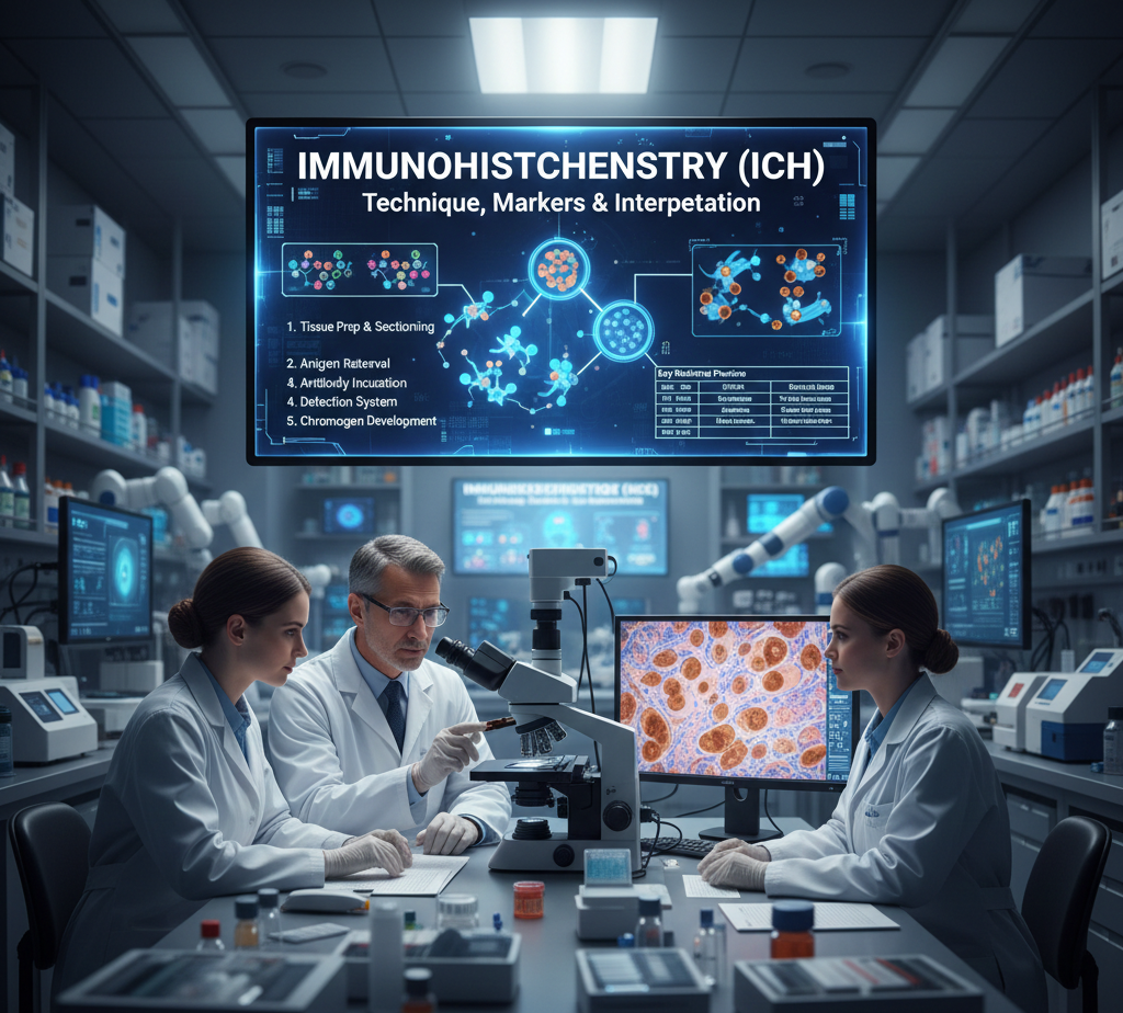
Tissue preparation is a process where the tissue is fixed in a liquid called formalin and then it is embedded in paraffin wax, which is also known as FFPE. This is a way to get the tissue ready for study. The tissue preparation process involves fixing the tissue and then embedding the tissue, in paraffin wax.
The tissue is cut into thin pieces, about 4 to 5 microns and then these pieces are put on glass slides. This is called sectioning of the tissue. The tissue is sectioned so it can be looked at closely.
Antigen Retrieval is a process where you use heat or special helpers like enzymes to make the antigens visible again. These antigens get hidden when a specimen is treated with formalin to preserve it. So when you use heat or enzymes it is like uncovering the antigens that were masked by the formalin. This way Antigen Retrieval helps us to see the antigens that were hidden which’s important, for studying them.
When we do Primary Antibody Incubation we put the Primary Antibody on and it sticks to the thing it is looking for which is the target antigen. The Primary Antibody is really good, at finding the target antigen and attaching to it.
The Detection System works like this: a special helper antibody that is connected to an enzyme, such, as HRP attaches itself to the antibody. This main antibody is what we call the antibody. The Detection System uses this antibody to get the job done.
Chromogen Development is a process where an enzyme works with a substance, like DAB and this makes a color that we can see. The enzyme and the substance, like DAB react together to produce this brown color. This is what happens in Chromogen Development.
Immunohistochemistry or IHC is not something that works the same for every situation. When doctors are trying to answer a question, about a patients health the pathologists will pick the type of stain that they think will help them find the answer. They have to choose between types of Immunohistochemistry stains.
Direct Immunohistochemistry or Direct IHC is a method where the primary antibody is labeled directly with a kind of dye called a fluorophore or an enzyme. This way of doing Direct IHC is quicker. It is not as good, at finding what we are looking for so it is less sensitive when we use Direct IHC.
Indirect IHC is the common method. It uses an antibody that does not have a label. This primary antibody is followed by an antibody that has a label. The signal gets stronger because one primary antibody can bind to secondary antibodies. This makes the Indirect IHC more sensitive. The Indirect IHC method is really good, at detecting things because of this.
Multiplex IHC is a way to find things in a tissue sample at the same time. It does this by using colors to show where different antigens are. This is really useful when we are trying to understand how cancer works, which is called oncology. Multiplex IHC is getting more important in this area because it helps us see antigens, in one tissue section.
The power of IHC is really in its collection of antibodies. When doctors look at tissues in pathology they use specific markers to figure out specific things about the tissue. They want to answer questions so they use specific markers, with IHC to do that. IHC is very useful because it has many antibodies that can help doctors find the answers they need.
Cytokeratins( CK7, CK20) Used to separate adenocarcinomas of different origins( e.g., lung vs. colon).
Thyroid Recap Factor- 1( TTF- 1) Specific for lung and thyroid lymphomas.
Desmin Indicates muscle isolation.
S- 100 Used for carcinoma, neural, and adipose towel excrescences.
CD3( T- cells) & CD20( B- cells) Essential for classifying tubercles.
Ki- 67 is a marker that shows how fast cells are growing. When Ki- 67 is high it means that the excrescences are veritably aggressive. The Ki- 67 marker is important because it helps us understand how serious the excrescences are. If a excrescence has a Ki- 67 position it’s likely to be a big problem. The Ki- 67 position is used to figure out how aggressive the excrescences are and what kind of treatment is demanded.
To really get what Immunohistochemistry or IHC can do for us in a setting it’s helpful to look at it next, to other ways we generally diagnose effects. We should compare IHC to individual tools that we use all the time. This will help us see how useful IHC really is.
point Immunohistochemistry( IHC) Flow Cytometry Molecular Testing( PCR/ NGS)
Sample Type Solid Towel( FFPE blocks) Fluid/ suspense( Blood, Bone Gist) Towel or Fluid( DNA/ RNA)
Spatial environment is really good when it preserves the armature.
On the hand Spatial environment is n’t present when cells are floating around in a suspense.
Spatial environment is also not there when the analysis is done on nucleic acids.
Protein Discovery yea( Direct visualization) Yes( face and intracellular) No( circular via DNA/ RNA)
Primary Use opinion, subtyping, prognostic labels Immunophenotyping, cell counts Mutation discovery, translocations
Turnaround Time 1 – 2 days Same day 3 – 10 days
Interpreting IHC is a skill that combines knowing about the body with understanding how antibodies work. For me interpreting IHC is where you really see if someone is good, at it. Interpreting IHC requires a lot of practice and knowledge of the body and antibodies.
Pathologists do n’t generally say commodity is just positive or negative. They frequently use scores that’re a little more complicated. For illustration the Allred score, which is used for bone cancer labels combines effects.
What chance of the cells are actually stained when you look at them? You have to give a Proportion Score from 0 to 5. This Proportion Score is important because it tells us how numerous cells are stained. The Proportion Score is like a measure of how important of the cellsre stained.
You need to suppose about what chance of the cellsre stained
also you give a Proportion Score from 0, to 5 grounded on that
The Proportion Score is used to figure out how numerous cells are stained.
Intensity Score( 0- 3) How strong is the stain( weak, moderate, strong)?
The total score is what determines how important commodity is in a setting for illustration whether or not someone has ER positivity. The total score is really what matters when it comes to figuring out significance like, in the case of ER positivity.
False Cons occasionally the biotin that’s formerly in our body like in the liver and order can get in the way of the discovery systems. This can beget staining that we do n’t want. The biotin that’s formerly in our body can bind to the discovery systems. This is what causes the problem. The discovery systems get confused by the biotin, in the liver and order. That’s why we get False Cons.
False Negatives If the towel is n’t fixed duly it can really mess up the antigens. This means that indeed if the protein we’re looking for is actually there we might not see any staining. The protein is present, in the towel. The poor obsession of the towel can make it feel like the protein is n’t there. This is a problem because it can give us results and we need to be careful when we’re fixing the towel so that we can find the protein if it’s really there.
Background staining is a problem because it’s caused by antibodies that bind to effects they are n’t supposed to. This can hide the signal that we’re trying to see. Background staining happens when antibodies stick to the effects and that makes it hard to get a clear picture of what’s really going on with the antibodies.
IHC is really important for figuring out what kind of excrescence someone has, especially when the excrescence does n’t look like it belongs to a group. When croakers look at a excrescence under a microscope and it just appears as a bunch of blue cells after using a standard H&E stain they use IHC to determine exactly where the excrescence came from. IHC helps croakers find out what type of IHC the excrescence is, which is pivotal for deciding the course of treatment, for the IHC and the excrescence.
For illustration, a excrescence in the lung could be
TTF- 1 positive Metastatic lung melanoma.
CDX- 2 positive Metastatic colon melanoma.
PAX- 8 positive Metastatic order melanoma.
The distinction of commodity makes a difference, in how we treat it. We’ve to use different styles to deal with the thing because of this distinction. The treatment protocols are different because of the distinction.
What’s the difference between IHC and Immunofluorescence( IF)?
Both of these styles use antibodies. The first one, which is called IHC uses a kind of coadjutor that makes a brown or blue color. We can see this color when we look at it with a microscope that uses regular light.The other system, which is called IF uses aides that glow in the dark. We need to use a kind of microscope to see these aides one that can show us glowing effects. IHC is what croakers generally use when they’re looking at apkins to figure out what’s wrong with someone.However, from a order, If is frequently used when croakers are trying to learn further about commodity or when they’re looking at a special kind of towel sample.
Immunohistochemistry or IHC can be done on kinds of towel. This is because IHC is a test that uses antibodies to find proteins in towel. IHC is generally performed on towel that has been taken out of the body. The towel is also treated with chemicals and cut into veritably thin slices. These slices are put on a glass slide. The antibodies are added to see if the proteins are present. IHC can be performed on towel from a vivisection or from towel that has been removed during surgery. So to answer the question IHC can be performed on types of towel including towel, from the skin, liver and other organs.
This thing generally works,. It works really well on towel that has been fixed with formalin and bedded in paraffin, which is called formalin- fixed paraffin- bedded or FFPE towel. People also use frozen towel but that requires special care when handling it. occasionally when bone towel is treated to remove calcium it can damage the antigens. That makes it hard to do commodity called IHC or Immunohistochemistry on the towel. IHC is still possible. It’s more delicate when the bone towel has been decalcified.
How long does an IHC stain take?
A single stain on a sample generally takes one to two days to get the results.
still when croakers run a lot of stains like ten, to twenty to understand a excrescence it can take several days.
This is especially true if the croakers need to do some work to make sure the tests are accurate.
Is IHC quantitative?
This thing is n’t fully measured. Digital pathology and image analysis software are trying to make it more measured. The mortal eye is still the stylish way to figure out what the stain really means for the case. The mortal eye is what we trust most when it comes to understanding the stain.
What are the limitations of IHC?
IHC is n’t suitable to find mutations or translocations on its own. still IHC can give us suggestions, about them. For illustration IHC can descry ALK staining in lung cancer. Ihc needs a good antibody to work duly. When croakers look at the results they’ve to use their own judgment because there are no standard rules to follow.
Spatial environment Immunohistochemistry or IHC for short is a way to see proteins in the towel and it keeps the towel structure complete so you can see where the proteins are, which is really important, for understanding what’s going on in the towel.
Diagnostic Triad It’s one of three pillars of pathology, alongside Histology( H&E) and Molecular testing.
Semi-Quantitative results are looked at in a way. TheSemi-Quantitative results are grounded on how commodity is stained and how strong the stain is. This meansSemi-Quantitative results are n’t just saying commodity is positive or negative.
Critical for Subtyping Essential for distinguishing between lymphomas, sarcomas, and tubercles, and for relating the point of origin for metastatic complaint.
Immunohistochemistry( IHC) islands the gap between the bitsy appearance of a cell and its molecular identity. By imaging protein expression within the towel environment, IHC provides pathologists with the precise data demanded to diagnose complex conditions and companion substantiated treatment plans. As oncology moves toward targeted curatives, the part of IHC in relating remedial biomarkers will only continue to grow.