Physical Address
304 North Cardinal St.
Dorchester Center, MA 02124
Physical Address
304 North Cardinal St.
Dorchester Center, MA 02124

Microbiology is often misrepresented as a science of passive observation—a discipline where one merely watches through lenses. However, the modern microbiology laboratory is more akin to a sophisticated engineering world. It is a facility where the laws of physics are manipulated to create sterility, chemical dyes act as structural stress tests for cell walls, and the evolutionary velocity of antibiotic resistance is recorded in real-time. Sterilization, Staining, Culturing, and Identification. This article moves beyond textbook definitions to analyze the thermodynamic and biochemical realities of these protocols.
The validity of any microbiology experiment rests on a binary condition: either the environment is sterile, or the data is invalid. There is no middle ground. Most reported contamination incidents stem from a failure to distinguish between sterilization (total destruction) and disinfection (reduction).
Sterilization refers to the absolute destruction of all microbial life, including the most resistant biological structures known: bacterial endospores (e.g., Bacillus and Clostridium). Disinfection, conversely, merely reduces pathogenic agents to a safe handling level but does not guarantee spore destruction. To achieve sterility, we must overcome the thermodynamic stability of microbial proteins.
| Method | Agent | Critical Parameter | Mechanism of Action | Limitations |
| Moist Heat | Saturated Steam | $121^\circ \mathrm{C}$ @ 15 psi | Coagulation & Hydrolysis | Cannot use on heat-sensitive plastics. |
| Dry Heat | Oxidative Air | $160^\circ \mathrm{C}$ for 2 hrs | Oxidation (Burning) | Poor heat transfer; requires long duration. |
| Filtration | Membrane | $0.22\ \mu m$ pore size | Exclusion (Physical) | Does not remove viruses or mycoplasmas. |
The autoclave is the thermodynamic engine of the lab. It utilizes saturated steam under pressure.
Mechanism: Moist heat kills via the coagulation of proteins. Water molecules disrupt hydrogen bonds at lower temperatures than dry air can.
The Steam Quality Variable: A critical, often overlooked operational data point is “steam quality.” Proper sterilization requires 97% saturated steam. If steam is too “wet” (>3% water), it causes localized cooling. If it is superheated (too dry), it acts like hot air—an insulator—and fails to kill spores even at $121^\circ \mathrm{C}$.
Used for moisture-resistant items like glassware, oils, and powders.
Mechanism: Oxidation. Essentially, the microorganism is incinerated to ash.
Protocol: Because dry air transfers heat inefficiently, it requires higher temperatures ($160^\circ \mathrm{C}$) for significantly longer periods (2 hours).
Used for heat-labile liquids such as antibiotics and vitamin solutions.
Mechanism: Exclusion. It uses a physical barrier, typically a $0.22\ \mu m$ membrane.
Analytical Note: Filtration is not absolute sterilization. Viruses ($0.02–0.1\ \mu m$) and Mycoplasmas lack cell walls and are small enough to pass through standard filters.
Invented by Hans Christian Gram in 1884, the Gram stain remains the single most critical diagnostic test. It is not just a coloring method; it is a structural stress test of the bacterial cell wall.
The reaction depends on the thickness and cross-linking density of the Peptidoglycan layer.
Gram-Positive Bacteria: Possess a massive, mesh-like peptidoglycan structure (20–80 nm thick) interlaced with teichoic acids. This structure is robust and resists dehydration.
Gram-Negative Bacteria: Possess a very thin peptidoglycan layer (2–7 nm) surrounded by a lipid-rich Outer Membrane containing lipopolysaccharides (LPS). This membrane is soluble in organic solvents.
The precision of the Gram stain hinges entirely on the third step: Decolorization.
Primary Stain (Crystal Violet): Dissociates into $CV^+$ ions that stain all cells purple.
Mordant (Gram’s Iodine): Forms a large Crystal Violet-Iodine (CV-I) Complex. This molecule is physically too large to easily escape the cell matrix.
Decolorizer (95% Ethanol/Acetone):
In Gram-Positives: Alcohol dehydrates the thick wall, shrinking pores and locking the CV-I complex inside. Result: Purple.
In Gram-Negatives: Alcohol dissolves the outer membrane and fails to dehydrate the thin wall enough to trap the dye. The complex washes out. Result: Colorless.
Counterstain (Safranin): Stains the colorless Gram-negatives pink.
The Age Factor: Gram-positive bacteria older than 24 hours undergo autolysis. Enzymes digest the cell wall, causing it to lose its holding power. A Staphylococcus (G+) may appear pink (G-), leading to a false diagnosis.
Over-Decolorization: Exposing the slide to alcohol for just 10 seconds too long can strip the dye from Gram-positive cells.
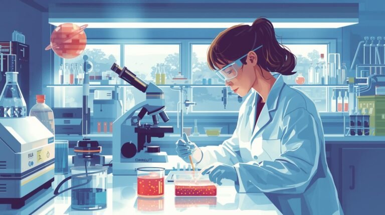
If Gram staining is the “fingerprint,” Culture and Sensitivity (C&S) is the interrogation. This process propagates the organism to identify it and determines which “biological weapons” (antibiotics) will destroy it.
We use specific biochemical formulations to filter biological “noise.”
Enriched Media (e.g., Blood Agar): The baseline. It allows for the visual assessment of Hemolysis (lysis of red blood cells).
Beta-hemolysis: Complete clearing (e.g., Streptococcus pyogenes).
Alpha-hemolysis: Partial greening (e.g., Streptococcus pneumoniae).
Selective/Differential Media (e.g., MacConkey Agar): The primary tool for gut bacteria.
Selectivity: Contains Bile Salts and Crystal Violet to kill Gram-positives.
Differentiation: Contains Lactose. Fermenters release acid, turning colonies pink (E. coli). Non-fermenters remain colorless (Salmonella).
Once isolated, the pathogen is tested against antimicrobials.
A qualitative method where antibiotic disks are placed on a bacterial lawn. As the drug diffuses, it creates a Zone of Inhibition—an area where concentration is sufficient to stop growth.
Limitation: It is binary. It categorizes bacteria as Sensitive, Intermediate, or Resistant, but does not provide dosage data.
This is the quantitative gold standard. It determines the lowest concentration ($\mu g/mL$) of a drug required to inhibit visible growth.
Clinical Relevance: If the MIC is $2\ \mu g/mL$, but the maximum safe drug level in the patient’s blood is only $1\ \mu g/mL$, the treatment will fail, even if the bacteria are technically “sensitive” in the petri dish.
The microbiology laboratory is the intersection of biology, chemistry, and physics. From the thermodynamics of the autoclave to the differential permeability of the cell wall, every protocol is a calculated scientific query. With antimicrobial resistance rising, understanding these mechanical underpinnings is no longer just academic—it is the frontline of the global health battle.